Focused echocardiography in the potential cardiac donor
This guidance is for doctors and advanced critical care practitioners carrying out a focused bedside echocardiography (echo) scan to assess a potential cardiac donor.
If you are fully accredited, please scan and report the echo according to British Society of Echocardiography (BSE) standards.
If you are focused echo accredited (Fusic Heart or Level I BSE), please scan and report according to your accreditation standards.
Once you have captured the images, it is important to promptly transfer them to NHS Blood and Transplant for expert assessment.
Please upload your image files to PACS and then arrange for them to be sent via the Image Exchange Portal (IEP) to "NHS Blood & Transplant". You may need to contact your hospital's PACS team to facilitate this.
Remember to adhere strictly to your scope of competence, and do not report findings beyond your expertise.

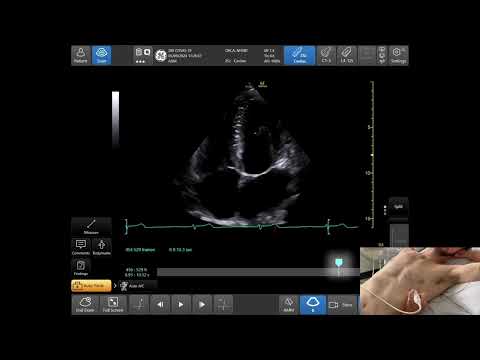
Training video for the donor heart transthoracic echo assessment
Watch this training video to guide you through the views and measurements set out in the guidance.
This video was created in collaboration with ORACLE Education – a free interactive online interface for radiology education hosted by The Postgraduate Virtual Learning Environment (@pgvle).
Views
Consider recording the following 18 views if possible and transfer images to the transplant centre.
Remote image review is essential in all cases.
See the measurement guide for all views marked with an asterisk (*).
Parasternal long axis
- 2D
- Colour over aortic valve*
- Colour over mitral valve*
- Measure*:
- Intraventricular septum thickness
- Posterior wall thickness
- End diastolic LV diameter
Parasternal short axis
- 2D Aortic level
- Colour over tricuspid*
- Colour over pulmonary valve*
- 2D Mitral level
- 2D Papillary muscle level
- 2D Apical level
Apical 4 Chamber
- 2D
- Colour over mitral valve*
- Colour over tricuspid valve*
- Measure*: RV basal diameter
Apical 5 Chamber
- 2D
- Colour over aortic valve*
Subcostal
- 2D
- Colour over inter-atrial septum*
Reporting
If you feel able to, please comment on the following:
- Inotrope/vasopressor level
- PEEP on ventilator
- LV function
- RV function
- Mitral valve
- Tricuspid valve
- Pulmonary valve
- Other (eg VSD/effusions)
- LV diameter (cm)
- LV septal wall thickness (cm)
- LV posterior wall thickness (cm)
- RV basal diameter (cm)
Please transfer the images to the transplant centre.
Advanced
If you are able to perform a complete BSE Level 2 Echo, this would be ideal.
Please record LVEF, regional wall abnormalities, RV function and any valvular abnormalities with quantification.
Many changes occur at end of life and do not necessarily mean transplantation is impossible – for example, regional wall motion abnormalities (RWMA).
Parasternal long axis measurements
Measure these parameters in diastole (when the LV is the biggest):
A: Intraventricular septum thickness
B: End diastolic LV diameter
C: Posterior wall thickness

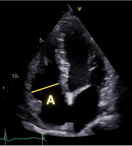
Apical 4 Chamber measurement
A: RV basal diameter in diastole (when RV is biggest)
Colour Nyquist limit
When taking colour images, ensure the colour scale Nyquist limit is set between 50 to 60cm/s.
A wide box to capture any valvular lesion is useful, but too wide and the image frame rate will reduce.

About this guidance
This guidance has been developed through a multidisciplinary working group, which was established to determine the key parameters needed for transplant teams to consider dispatching a retrieval team to assess suitable heart donors.
Many of the scans performed for donor heart assessment are done by those working towards or having completed focused echocardiography accreditation.
For this reason, we have outlined a pragmatic approach in this guidance. It focuses on the acquisition of essential imaging, even in situations where local expertise may not extend to the full interpretation of the images.
For background and context on the above guidance, please read these peer-reviewed publications:
- Akhtar W, Peck M, Miller A and others. 'NHS blood and transplant donor echocardiography standard to improve organ utilisation in heart transplantation' Journal of the Intensive Care Society 2025
- Akhtar W, Marshal L, Buglass H and others. 'A survey of United Kingdom intensive care echocardiography provision' Journal of the Intensive Care Society 2024: volume 25, issue 4, pages 407-409
- Akhtar W, Padukone A, Rowson R and others. 'A UK-wide prospective assessment of donor heart echocardiography pathway'. The British Journal of Cardiology 2024: volume 31, pages 136-8